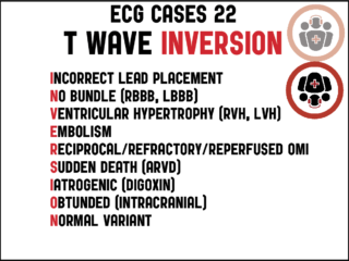
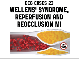
ECG Cases 23 – Wellens syndrome, reperfusion and reocclusion MI

Eight patients presented with potentially ischemic symptoms and T-wave inversions. Which had occlusion MI, which were reperfused and which were reoccluded? Jesse McLaren helps you discover the nuances of Wellens syndrome and T-wave inversions on this month's ECG Cases blog...

By |2021-07-13T12:27:47-04:00July 13th, 2021|Categories: Cardiology, ECG Cases, Emergency Medicine, Medical Specialty|Tags: , , , |0 Comments

ECG Cases 22: T-wave INVERSION mnemonic

The differential for T-wave INVERSION includes: Incorrect lead placement, No bundle (RBBB, LBBB), Ventricular hypertrophy (LVH, RVH), Embolism, Reciprocal/refractory/reperfused occlusion MI, Sudden death (ARVD), Iatrogenic (digoxin), Obtunded (eg SAH), and Normal variant. Jesse McLaren runs through 10 cases of patients who present to the ED who have T-wave inversions on their ECGs...

ECG Cases 21: Hyperacute T-waves and Occlusion MI

In this month's ECG Cases blog, Dr. Jesse MacLaren guides us through 10 cases of patients whose ECGs show tall T-waves and gives us the tools to distinguish the hyperacute T-waves of occlusion MI from other causes of tall T-waves....

ECG Cases 20 – Approach to Bradycardia and the BRADI Mnemonic

In this ECG Cases we review 10 patients who presented with bradycardia, introduce an approach to reversible causes using the BRADI mnemonic and guide you through how to use the ECG to guide management...

ECG Cases 17 – ST ELEVATIONS mnemonic and Occlusion MI

In this ECG Cases blog we look at 10 patients who presented with ST elevation, and review the differential diagnosis of ST elevation using the ELEVATIONS mnemonic. Which had occlusion MI?

ECG Cases 14: Q-wave and Occlusion MI

9 patients presented with potentially ischemic symptoms and Q-waves. Which had Occlusion MI? What is the differential diagnosis of Q-waves? How can one distinguish MI vs LV aneurysm on ECG?...

By |2020-10-13T12:31:07-04:00October 13th, 2020|Categories: Cardiology, ECG Cases, EM Cases, Emergency Medicine, Medical Specialty|Tags: , , , |0 Comments

ECG Cases 13: LVH and Occlusion MI

LVH produces secondary repolarization abnormalities that can mimic STEMI. Signs of occlusion MI in patients with LVH include: new Q waves/loss of R waves, disproportionate and dynamic ST elevation (or ST depression from posterior MI), and hyperacute T waves. In this ECG Cases blog we look at 6 patients who presented with potentially ischemic symptoms and LVH on their ECG. Which had an acute coronary occlusion?

By |2022-08-14T22:08:57-04:00September 15th, 2020|Categories: Cardiology, ECG Cases, EM Cases, Medical Specialty|Tags: , , , |0 Comments

ECG Cases 11: LBBB + Occlusion MI

In ECG Cases 11 - LBBB + Occlusion MI Jesse McLaren runs through 5 patients who present with potentially ischemic symptoms and LBBB and explains the evolution of decision making for cath lab activation criteria. The two most important points are that we should always use the Smith-modified Sgarbossa Criteria to identify occlusion MI in the patient with ischemic symptoms and LBBB, whether old or new, and that those with refractory ischemia and hemodynamic instability from suspected occlusion MI require cath lab activation regardless of the ECG....

Donate Subscribe
Go to Top