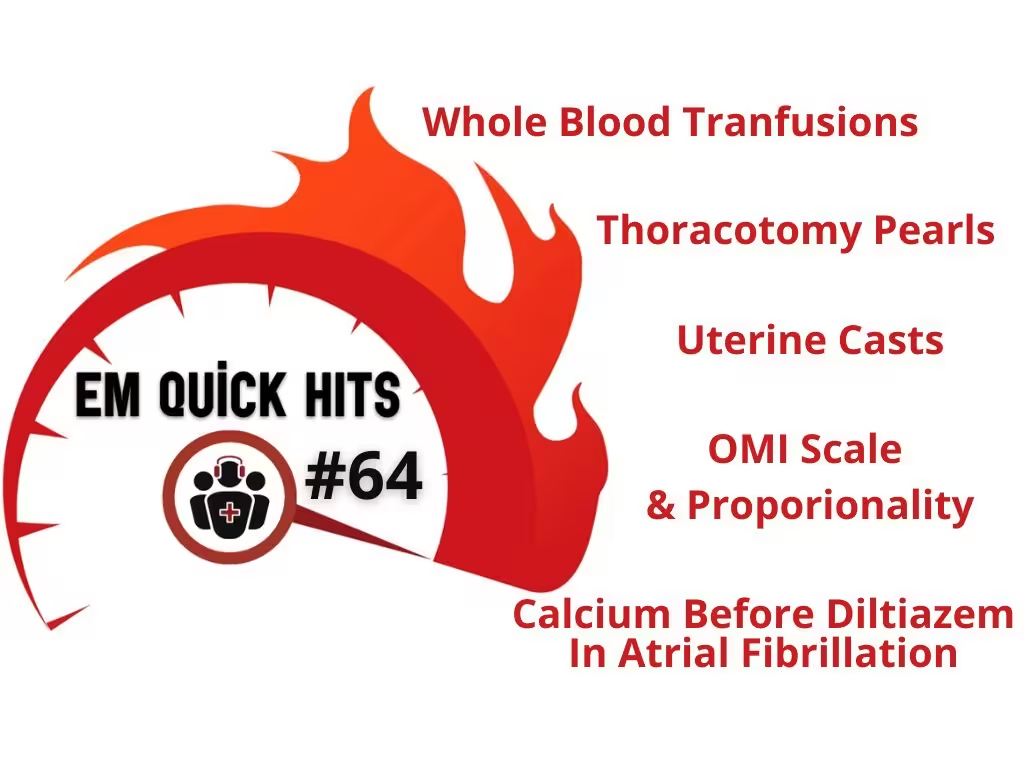
Topics in this EM Quick Hits podcast
Zafar Qasim & Andrew Petrosoniak on whole blood transfusion in trauma (1:32)
Justin Morgenstern on calcium pre-treatment to prevent diltiazem-induced hypotension (29:57)
Kiran Rikhraj on dynamic LV outflow tract obstruction (36:35)
Anand Swaminathan on resuscitative thoracotomy (42:35)
Andrew Tagg on uterine casts (48:22)
Jesse McLaren on scale & proportionality in occlusion MI ECG interpretation (53:38)
Podcast production, editing and sound design by Anton Helman
Podcast content, written summary & blog post by Brandon Ng, edited by Anton Helman, April, 2025
Cite this podcast as: Helman, A. Swaminathan, A. Qasim, Z, M. Petrosoniak, A. Rikhraj, K, Tagg, A. Morgenstern J. EM Quick Hits 64 Whole Blood Transfusions, Calcium Before Diltiazem in Afib, Thoracotomy Pearls, Uterine Casts, OMI Scale & Proportionality. Emergency Medicine Cases. April, 2025 https://stg-emergencymedicinecases-emcstaging.kinsta.cloud/em-quick-hits-month-year/. Accessed July 2, 2025.
Whole blood transfusions in trauma resuscitation: Better than component therapy?
Current Practice: Most centers use component therapy (RBCs, plasma, platelets) with a 1:1:1 ratio based on evidence from the PROPPR trial (Holcomb et al. 2015) rather than whole blood tranfusions. However, there is a recent shift in some U.S. centers towards adopting whole blood therapy in trauma resuscitation.
Benefits of whole blood transfusion therapy
(Typically refers to cold-stored low-titer O whole blood in civilian practices)
- Simplicity: Simpler to administer and logistically less complex than separate components, with universal compatibility and no need to balance component ratios.
- Evidence: Observational studies in military settings show improved early survival with warm fresh whole blood compared to component therapy (Gurney et al. 2020, Shackelford et al. 2021).
- Physiologic advantages: Provides higher hematocrit, platelet counts, and clotting factor content in a whole blood unit when compared to equivalent component therapy.
- Safety: May have fewer transfusion-related complications.
Arguments against using whole blood transfusions, challenges to implementation
- Limited RCT Evidence: Robust RCT evidence is lacking to support widespread adoption; data to support whole blood as superior to component therapy is primarily observational.
- Rh Sensitization Risk: Potential for alloimmunization in Rh- women of childbearing age receiving Rh+ whole blood, posing a risk for hemolytic disease of the newborn in future pregnancies.
- Logistics: Supply is very dependent on donor availability, which can fluctuate.
- Storage: Platelet function declines over time in stored whole blood. Solutions include rotating stock to high-usage centers or converting near-expiry units to component products.
Read more about massive hemorrhage protocols in Ep 152 “The 7 Ts of Massive Hemorrhage Protocols”
How Can Whole Blood be Used in a Massive Hemorrhage Protocol?
- Example protocol from Dr. Qasim: Patients who present with an ABC score ≥2 (e.g., penetrating trauma, hypotension, +FAST, tachycardia) receive up to 4 units of whole blood before switching to component therapy.
- Observational studies suggest earlier is better: every minute delay beyond 14 mins may worsen survival outcomes (Torres et al. 2024).
- Whole blood is typically stopped after 4 units or when hemorrhage control is achieved. However, evidence is limited, and ongoing RCTs like TROOP trial may provide further guidance.
Recent observational studies and guidelines (e.g. Meizoso et al. 2024, van der Horst et al. 2023, Torres et al. 2023) support whole blood in reducing early 24-hour mortality, through there is limited data on long-term outcomes.
Injury-specific data (e.g. concomitant traumatic brain injury, penetrating vs. blunt trauma) are sparse. Further studies focusing on injury subgroups may help clarify which patients benefit most from whole blood.
Bottom line => Whole blood is logistically simpler, physiologically superior in theory, and possibly associated with improved early 24-hour survival. Further RCTs are needed to determine its long-term safety and outcomes.
- Holcomb JB, Tilley BC, Baraniuk S, Fox EE, Wade CE, Podbielski JM, del Junco DJ, Brasel KJ, Bulger EM, Callcut RA, Cohen MJ, Cotton BA, Fabian TC, Inaba K, Kerby JD, Muskat P, O’Keeffe T, Rizoli S, Robinson BR, Scalea TM, Schreiber MA, Stein DM, Weinberg JA, Callum JL, Hess JR, Matijevic N, Miller CN, Pittet JF, Hoyt DB, Pearson GD, Leroux B, van Belle G; PROPPR Study Group. Transfusion of plasma, platelets, and red blood cells in a 1:1:1 vs a 1:1:2 ratio and mortality in patients with severe trauma: the PROPPR randomized clinical trial. JAMA. 2015 Feb 3;313(5):471-82. doi: 10.1001/jama.2015.12. PMID: 25647203; PMCID: PMC4374744.
- Gurney J, Staudt A, Cap A, Shackelford S, Mann-Salinas E, Le T, Nessen S, Spinella P. Improved survival in critically injured combat casualties treated with fresh whole blood by forward surgical teams in Afghanistan. Transfusion. 2020 Jun;60 Suppl 3:S180-S188. doi: 10.1111/trf.15767. Epub 2020 Jun 3. PMID: 32491216.
- Shackelford SA, Gurney JM, Taylor AL, Keenan S, Corley JB, Cunningham CW, Drew BG, Jensen SD, Kotwal RS, Montgomery HR, Nance ET, Remley MA, Cap AP; Joint Trauma System Defense Committee on Trauma; Armed Services Blood Program. Joint Trauma System, Defense Committee on Trauma, and Armed Services Blood Program consensus statement on whole blood. Transfusion. 2021 Jul;61 Suppl 1:S333-S335. doi: 10.1111/trf.16454. PMID: 34269445.
- Torres CM, Kenzik KM, Saillant NN, Scantling DR, Sanchez SE, Brahmbhatt TS, Dechert TA, Sakran JV. Timing to First Whole Blood Transfusion and Survival Following Severe Hemorrhage in Trauma Patients. JAMA Surg. 2024 Apr 1;159(4):374-381. doi: 10.1001/jamasurg.2023.7178. Erratum in: JAMA Surg. 2024 Apr 1;159(4):470. doi: 10.1001/jamasurg.2024.0324. PMID: 38294820; PMCID: PMC10831629.
- Meizoso JP, Cotton BA, Lawless RA, Kodadek LM, Lynde JM, Russell N, Gaspich J, Maung A, Anderson C, Reynolds JM, Haines KL, Kasotakis G, Freeman JJ. Whole blood resuscitation for injured patients requiring transfusion: A systematic review, meta-analysis, and practice management guideline from the Eastern Association for the Surgery of Trauma. J Trauma Acute Care Surg. 2024 Sep 1;97(3):460-470. doi: 10.1097/TA.0000000000004327. Epub 2024 Mar 27. PMID: 38531812.
- van der Horst RA, Rijnhout TWH, Noorman F, Borger van der Burg BLS, van Waes OJF, Verhofstad MHJ, Hoencamp R. Whole blood transfusion in the treatment of acute hemorrhage, a systematic review and meta-analysis. J Trauma Acute Care Surg. 2023 Aug 1;95(2):256-266. doi: 10.1097/TA.0000000000004000. Epub 2023 May 1. PMID: 37125904.
- Torres CM, Kent A, Scantling D, Joseph B, Haut ER, Sakran JV. Association of Whole Blood With Survival Among Patients Presenting With Severe Hemorrhage in US and Canadian Adult Civilian Trauma Centers. JAMA Surg. 2023 May 1;158(5):532-540. doi: 10.1001/jamasurg.2022.6978. PMID: 36652255; PMCID: PMC9857728.
- Cotton BA, Podbielski J, Camp E, Welch T, del Junco D, Bai Y, Hobbs R, Scroggins J, Hartwell B, Kozar RA, Wade CE, Holcomb JB; Early Whole Blood Investigators. A randomized controlled pilot trial of modified whole blood versus component therapy in severely injured patients requiring large volume transfusions. Ann Surg. 2013 Oct;258(4):527-32; discussion 532-3. doi: 10.1097/SLA.0b013e3182a4ffa0. Erratum in: Ann Surg. 2014 Jul;260(1):178. PMID: 23979267.
Calcium for prevention of diltiazem-induced hypotension in management of rapid atrial fibrillation
The Paper: Reducing diltiazem-related hypotension in atrial fibrillation: Role of pretreatment intravenous calcium by Az et al. AJEM 2025.
This single-center randomized controlled trial suggests that IV calcium pretreatment may prevent diltiazem-induced hypotension in patients with Afib/Aflutter with HR >120BPM.
- P: Adult patients with Afib/Aflutter and HR >120 BPM
- I: 2 intervention groups: 90mg IV calcium chloride (C90D) OR 180mg IV calcium chloride (C180D) pretreatment before IV diltiazem
- C: IV NS placebo pretreatment before IV diltiazem
- O: SBP at 5 min: Significantly lower in the control group compared to C90D/C180D groups. SBP at 10 min: Significantly higher in C180D group compared to control/C90D groups. SBP at 15 min: Significantly higher in both C90D/C180D compared to the controlled group. HR: significantly lower in the control/C90D group compared to the C180D group at 10 and 15 min. No significant differences in adverse events between groups. Overall, there is a decrease in SBP in both the control and C90D group, but not in the C180D group by 15 minutes.
Limitations of This Study:
- Hypotension from diltiazem was not clinically significant: Despite SBP differences between groups, all groups maintained SBP >115 at any time, and there were no statistical differences in hypotention rates.
- Rapid push dosing of 0.25mg/kg IV diltiazem likely exaggerated any hypotensive effect of diltiazem as compared to typical clinical practice where it is usually given at a slower rate (e.g. over 15-20 min).
- Calcium chloride is a caustic agent which may lead to harms from extravasation (e.g. necrosis). However, the study does not have sufficient power to measure these rare but important adverse outcomes. Calcium gluconate is generally preferred.
Bottom line => Results of the trial do not demonstrate clinically significant benefits of calcium pretreatment. Calcium should not be given routinely pending further evidence. Consider calcium gluconate for high-risk or borderline hypotensive patients.
- Az A, Sogut O, Dogan Y, Akdemir T, Ergenc H, Umit TB, Celik AF, Armagan BN, Bilici E, Cakmak S. Reducing diltiazem-related hypotension in atrial fibrillation: Role of pretreatment intravenous calcium. Am J Emerg Med. 2025 Feb;88:23-28. doi: 10.1016/j.ajem.2024.11.033. Epub 2024 Nov 17. PMID: 39577214.
Dynamic Left Ventricular Outflow Tract Obstruction (LVOTO): An important consideration in pressor-resistant shock
What is Dynamic Left Ventricular Outflow Tract Obstruction?
- Pathophysiology: fast-flowing blood flows through the LV outflow tract and creates a negative pressure effect on the mitral valve, causing the mitral valve be pulled into the LVOT and subsequently obstruct the LVOT (this phenomenon is referred to as systolic anterior motion)
- LVOT obstruction leads to profound obstructive shock
- LVOT can occur in the following conditions:
- Distributive shock with volume depletion
- Hypertrophic obstructive cardiomyopathy (HOCM)
- Hypertensive cardiomyopathy
- Takotsubo cardiomyopathy
- Left anterior descending artery (LAD) OMI with apical hypokinesis
- Cor pulmonale
How to identify Dynamic LVOTO?
- Think dynamic LVOTO when a tachycardic patient worsens with escalating doses of ionotropes and vasopressors and has a clinical picture of obstructive shock after consideration for immediate life threats of causing obstructive shock such as PE, cardiac tamponade and tension pneumothorax.
- Hyperdynamic LV on PoCUS. The hallmark PoCUS finding is a high-velocity, late-peaking continuous-wave Doppler signal in the LV outflow tract (referred to as “dagger-shaped”). (see video below).
- Clues of anatomical causes on PoCUS including hypertrophic cardiomyopathy, left ventricular hypertrophy, or sigmoid septum.
- If no anatomical causes identified, consider physiologic conditions that can cause dynamic LVOTO via increasing LV preload, decreasing LV afterload, increasing LV contractility: Hypovolemia, sepsis, anaphylaxis, use of inotropes, significant tachycardia.
- Doppler ultrasound may confirm diagnosis (see video below)
Management:
- Treat underlying cause (e.g. massive hemorrhage protocol, antibiotics, steroids)
- Increase LV preload: IV fluids, pure vasoconstrictors (e.g. vasopressin, phenylephrine)
- Increase LV afterload: stop nitrates/hydralazine (afterload reducing medications), start phenylephrine/vasopressin. Beware of norepinephrine (causes tachycardia, monitor HR if used)
- Reduce LV contractility: stop inotropes (e.g. epinephrine, dobutamine, milrinone)
- Decrease heart rate: cautiously use short-acting beta-blocker (e.g., esmolol) if persistent shock
Bottom line => Think dynamic LVOTO in shock patients who worsen with inotropes/vasopressors after other immediate life threats of obstructive shock have been considered. Stop inotropes, slow HR, and improve afterload/preload.
- Evans JS, Huang SJ, McLean AS, Nalos M. Left ventricular outflow tract obstruction-be prepared! Anaesth Intensive Care. 2017 Jan;45(1):12-20. doi: 10.1177/0310057X1704500103. PMID: 28072930.
- Sobczyk D. Dynamic left ventricular outflow tract obstruction: underestimated cause of hypotension and hemodynamic instability. J Ultrason. 2014 Dec;14(59):421-7. doi: 10.15557/JoU.2014.0044. Epub 2014 Dec 30. PMID: 26674265; PMCID: PMC4579722.
Resuscitative thoracotomy pearls and pitfalls
Indications for resuscitative thoracotomy
- Blunt trauma patients: consider only if arrest occurs in the ED – resuscitative thoracotomy unlikely to play a significant role in traumatic OHCA due to low survival rates
- Penetrating trauma with current or recent signs of life (present vital signs)
- Presence of pericardial effusion or cardiac activity on FAST exam
Tips On Achieving a Successful Resuscitative Thoracotomy:
- Position patient far left on bed, left arm overhead to provide access to the left side of the chest
- Anticipate the need for massive transfusion, calcium, TXA
- Prepare tools to address potential cardiac injury: stapler, 0-silk, needle driver, foley catheter, internal paddles (5-20J), intracardiac epinephrine
- Provide internal cardiac massage during cardiac repair using a rolling technique, not just squeezing
- At minimum do a finger thoracotomy of the right chest/place a chest tube in case of right-sided injuries. If injuries present → consider clamshell thoracotomy
- Closed chest compressions have no role in a traumatic arrest as it does not address common causes (e.g. pneumothorax, tamponade, exsanguination) – do not let it delay thoracotomy
Short thoracotomy video on cadaver: https://www.youtube.com/watch?v=A57ZB_J4FuY
Deeper dive thoracotomy video demonstration:
- Moore EE, Knudson MM, Burlew CC, Inaba K, Dicker RA, Biffl WL, Malhotra AK, Schreiber MA, Browder TD, Coimbra R, Gonzalez EA, Meredith JW, Livingston DH, Kaups KL; WTA Study Group. Defining the limits of resuscitative emergency department thoracotomy: a contemporary Western Trauma Association perspective. J Trauma. 2011 Feb;70(2):334-9.
- Hunt PA, Greaves I, Owens WA. Emergency thoracotomy in thoracic trauma-a review. Injury. 2006 Jan;37(1):1-19
- Lorenz HP, Steinmetz B, Lieberman J, Schecoter WP, Macho JR. Emergency thoracotomy: survival correlates with physiologic status. J Trauma. 1992 Jun;32(6):780-5; discussion 785-8.
- Khorsandi M, Skouras C, Shah R. Is there any role for resuscitative emergency department thoracotomy in blunt trauma? Interact Cardiovasc Thorac Surg. 2013 Apr;16(4):509-16.
- Slessor D, Hunter S. To be blunt: are we wasting our time? Emergency department thoracotomy following blunt trauma: a systematic review and meta-analysis. Ann Emerg Med. 2015 Mar;65(3):297-307.e16.
- DuBose J, Fabian T, Bee T, Moore LJ, Holcomb JB, Brenner M, Skarupa D, Inaba K, Rasmussen TE, Turay D, Scalea TM., AAST AORTA Study Group. Contemporary Utilization of Resuscitative Thoracotomy: Results From the AAST Aortic Occlusion for Resuscitation in Trauma and Acute Care Surgery (AORTA) Multicenter Registry. Shock. 2018 Oct;50(4):414-420.
Uterine casts: An important diagnosis mistaken as an abortion
The vignette highlights a 15-year-old girl presenting to the ED after having passed what appeared like a tiny uterus which was, in fact, a uterine cast.
What is a Uterine Cast?
- It is a rare but benign occurrence where the entire endometrial lining sheds in one piece
- Often mistaken for fetal abortion or tumor
- The exact cause is not fully understood, although abrupt progesterone withdrawal from the absence of egg fertilization and implantation likely plays a role
- Usually occurs during heavy or painful periods
- The passing of the large mass through the cervical os causes severe pain
Management of uterine casts
- Do not investigate
- Rule out pregnancy (b-HCG)
- Offer reassurance and pain control
Bottom line => Don’t over-investigate uterine casts. Provide reassurance to patients, as uterine casts are benign.
More on uterine casts at Don’t Forget the Bubbles
- Lankester E, Justice TD, Sachedina A, Rosenbaum DG. Decidual cast: a rare cause of genital tract obstruction. Pediatr Radiol. 2024 Aug;54(9):1549-1552. doi: 10.1007/s00247-024-05955-z. Epub 2024 May 24. PMID: 38787524.
- . Uterine (decidual) Casts, Don’t Forget the Bubbles, 2021. Available at: https://doi.org/10.31440/DFTB.32416
Occlusion MI: Scale and Proportionality on ECG
STEMI criteria looks at ST elevation in isolation and ignores scale and proportionality, thereby limiting its ability to detect occlusion MI and leading to high rates false positives and false negatives.
Scale = the size of one object compared to a different object.
Proportionality = size of one part of an object relative to other parts of that same object (i.e compare ST changes relative to the proceeding QRS).
Steps to identifying ST and T wave abnormalities
- Start by characterizing QRS/depolarization using the HEARTS approach (Heart rate & rhythm; Electrical conduction, Axis, R-wave progression, Tall or Small voltages).
- Assess the pair of ST segment and T wave after the QRS
- If QRS is normal, we can directly look for primary ischemic ST elevation and hyperacute T waves
- However, we can expect secondary, discordant, and proportional ST changes with abnormally wide (e.g. LBBB) or abnormally tall (e.g. LVH) QRS complexes
- Use the Smith-modified Sgarbossa criteria (Excessive discordance: >25% ST elevation relative to preceding S wave) to look for superimposed primary ischemic ST changes in the context of abnormal QRS
Bottom line: Don’t measure ST elevation in isolation. Use proportionality and clinical context to better detect occlusion MI.
For case examples and discussion on scale and proportionality: ECG Cases 54 – The Art of Occlusion MI: Scale and Proportionality
Register for Dr. McLaren’s HEARTS ECG Course to master your ECG interpretation skills.
None of the authors have any conflicts of interest to declare


Leave A Comment