cardiology emergency medicine
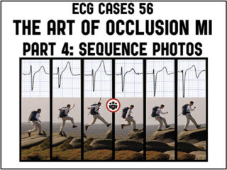
ECG Cases 56 – Art of Occlusion MI Part 4: Sequence Photos
In this month's ECG Cases Dr. McLaren explores how sequence photos help identify Occlusion MI. He illustrates through cases how hyperacute T waves and subtle ST elevation with reciprocal ST depression can provide an early snapshot of occlusion MI – and might remain the only sign of occlusion. How resolution of ischemic symptoms along with regional T wave inversion (or reciprocally tall anterior T waves) can indicate spontaneous reperfusion, while subacute and persisting symptoms with Q waves and T wave inversion indicate refractory occlusion. And how spontaneous reperfusion is at risk for reocclusion, with recurrence of ischemic symptoms accompanied by ST/T pseudonormalization and then hyperacute T waves and ST elevation.